Ophthalmology
The red eye, visual loss and eye emergencies.
Use as a study guide only. These notes are part of a free open-access medical education (FOAMed) project and may contain errors or outdated information. Always verify against current guidelines (e.g. eTG, RACGP, local health district policies) and reputable sources before applying anything to patient care. See the full disclaimer.
 OphthalmologyPDF · opens in a new tab
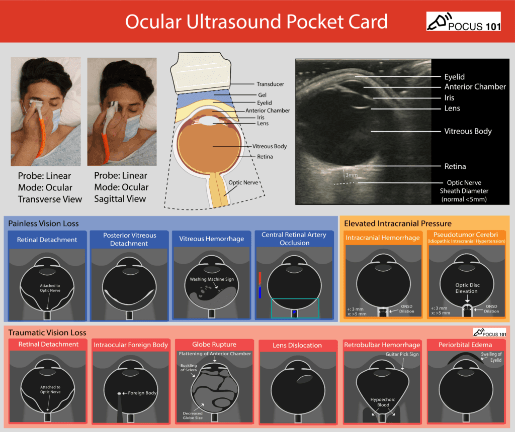 Ocular Ultrasound Pocket Card — PosterPDF · opens in a new tab
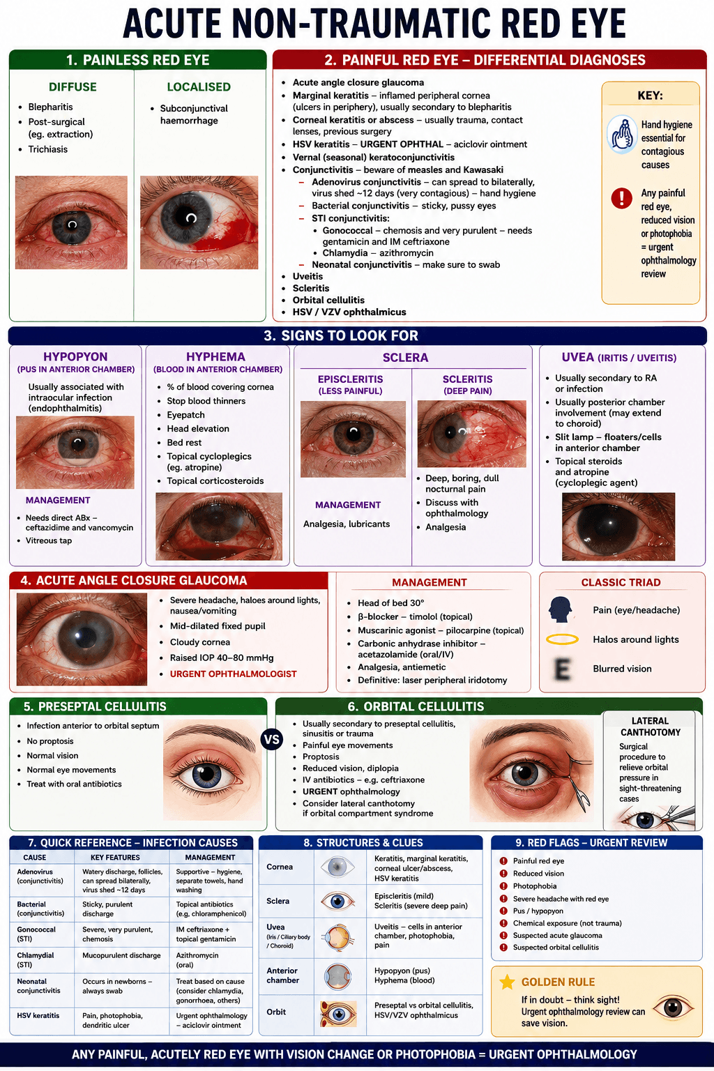 Acute Non-Traumatic Red Eye — PosterPDF · opens in a new tab
Clinical pearls & learnings
Tips and tricks collected over the years — a living list that grows with every rotation. Use as a study guide only and check current guidelines before acting on anything.
Assessment & referral
The ophthalmology referral in six steps
Opening line that gets attention: “Hi, I've got a [age] patient with acute painless vision loss in the right eye, VA 6/60, concern for retinal pathology.”
- 1. Visual acuity — best corrected (glasses/pinhole); the single most important number
- 2. Laterality — right / left / bilateral, acute vs chronic
- 3. Location — clock-face + distance from limbus or macula (e.g. “corneal ulcer at 2 o'clock, ~2mm from limbus”, “retinal tear at 10 o'clock, superotemporal”)
- 4. Key exam findings — anterior segment, pupil (size, reactivity, RAPD), fundoscopy (disc, retina, macula)
- 5. Red flags — state them explicitly
- 6. Intraocular pressure (tonometry) — unless penetrating trauma is suspected
- History must cover: trauma (high-velocity → think intraocular foreign body), past ocular history (glaucoma, uveitis, surgery), systemic disease (diabetes, autoimmune, GCA symptoms), medications (steroids, anticoagulants), contact lens use, recent surgery, immunosuppression.
- Red flags to mention explicitly: vision loss, painful eye, photophobia, trauma or chemical exposure, contact lens wear, RAPD, proptosis, severe headache (angle closure / GCA).
Ocular ultrasound: optic nerve sheath diameter
To assess for raised intracranial pressure on ocular ultrasound: measure 3mm posterior to the globe/retina, then measure the optic nerve sheath diameter at that point — >5mm suggests raised ICP.
Do not perform ocular ultrasound if globe rupture or penetrating injury is suspected — pressure on the globe can extrude intraocular contents.
Eye trauma & emergencies
Penetrating eye injury
If penetrating trauma is suspected: do NOT instil eye drops and do NOT measure pressure. Shield the eye (no patching/pressure) and refer urgently.
- Seidel test: fluorescein streaming away from a leak like a waterfall (the “waterfall sign”) confirms globe perforation.
- High-velocity mechanism (grinding, hammering) → CT orbits for intraocular foreign body.
Lateral canthotomy is not the treatment for a suspected open (penetrating/ruptured) globe — it treats a different problem (orbital compartment syndrome from retrobulbar haemorrhage, below). Cutting into a genuinely open globe risks worsening the injury; shield it and get urgent ophthalmology instead.
Orbital compartment syndrome
- Recognise: RAPD, reduced visual acuity, raised IOP, proptosis (typically after trauma with retrobulbar haemorrhage).
- Treat: urgent lateral canthotomy + cantholysis of the inferior tendon — sight-saving and time-critical.
- Practical setup: procedural sedation (e.g. ketamine), patient supine, gauze ready.
Hyphaema
- Blood in the anterior chamber, usually after blunt trauma.
- Manage with head elevation, eye protection (shield), and avoiding anticoagulants/antiplatelets.
- Monitor for raised intraocular pressure and for rebleeding (classically days 3–5).
Welder's arc / flash burns (photokeratitis)
Intensely painful but self-limiting. Treat with lubricating ointment and chloramphenicol ointment/drops for prophylaxis, plus oral analgesia — usually heals within 24–72 hours.
Retinitis pigmentosa & syndromes
RP and its associated syndromes
Retinitis pigmentosa (genetic photoreceptor dystrophy) = tunnel vision + night blindness (nyctalopia), with bone-spicule pigmentation on fundoscopy. If RP features appear with other systems involved, think of a syndrome:
| Syndrome | Clue | Distinguishing features |
|---|---|---|
| Usher syndrome | RP + deafness | Nyctalopia, peripheral vision loss, bone spicules + sensorineural hearing loss |
| Bardet–Biedl syndrome | RP + obesity | Polydactyly, learning difficulties, renal impairment |
| Refsum disease | Metabolic (excess phytanic acid) | Nyctalopia, anosmia, peripheral neuropathy, cerebellar signs |
| Kearns–Sayre syndrome | Mitochondrial, onset <20 years | Pigmentary retinopathy, progressive ophthalmoplegia, cardiac conduction defects (needs cardiology surveillance) |
The red, painful eye
Differentials not to miss
- Acute angle-closure glaucoma — severe pain, headache, haloes, fixed mid-dilated pupil.
- Uveitis — photophobia, cells/flare ± hypopyon; screen for systemic associations.
- Trauma — corneal abrasion, foreign body.
- Infection — keratitis (especially contact lens wearers), gonococcal conjunctivitis (hyperacute, sight-threatening).
- Optic neuritis — pain on eye movement, RAPD, reduced colour vision; think MS.
- Orbital cellulitis — proptosis, painful/restricted eye movements, systemically unwell.
PMR / GCA steroid ladder
| Scenario | Steroid approach |
|---|---|
| PMR alone | Low-dose prednisone (e.g. 15mg daily) |
| GCA (no visual symptoms) | High-dose prednisone (e.g. 40–60mg daily) |
| GCA with ocular symptoms | IV pulse methylprednisolone — treat immediately, do not wait for the biopsy |