Dermatology
Rashes, lesions and describing what you see.
Use as a study guide only. These notes are part of a free open-access medical education (FOAMed) project and may contain errors or outdated information. Always verify against current guidelines (e.g. eTG, RACGP, local health district policies) and reputable sources before applying anything to patient care. See the full disclaimer.
 DermatologyPDF · opens in a new tab
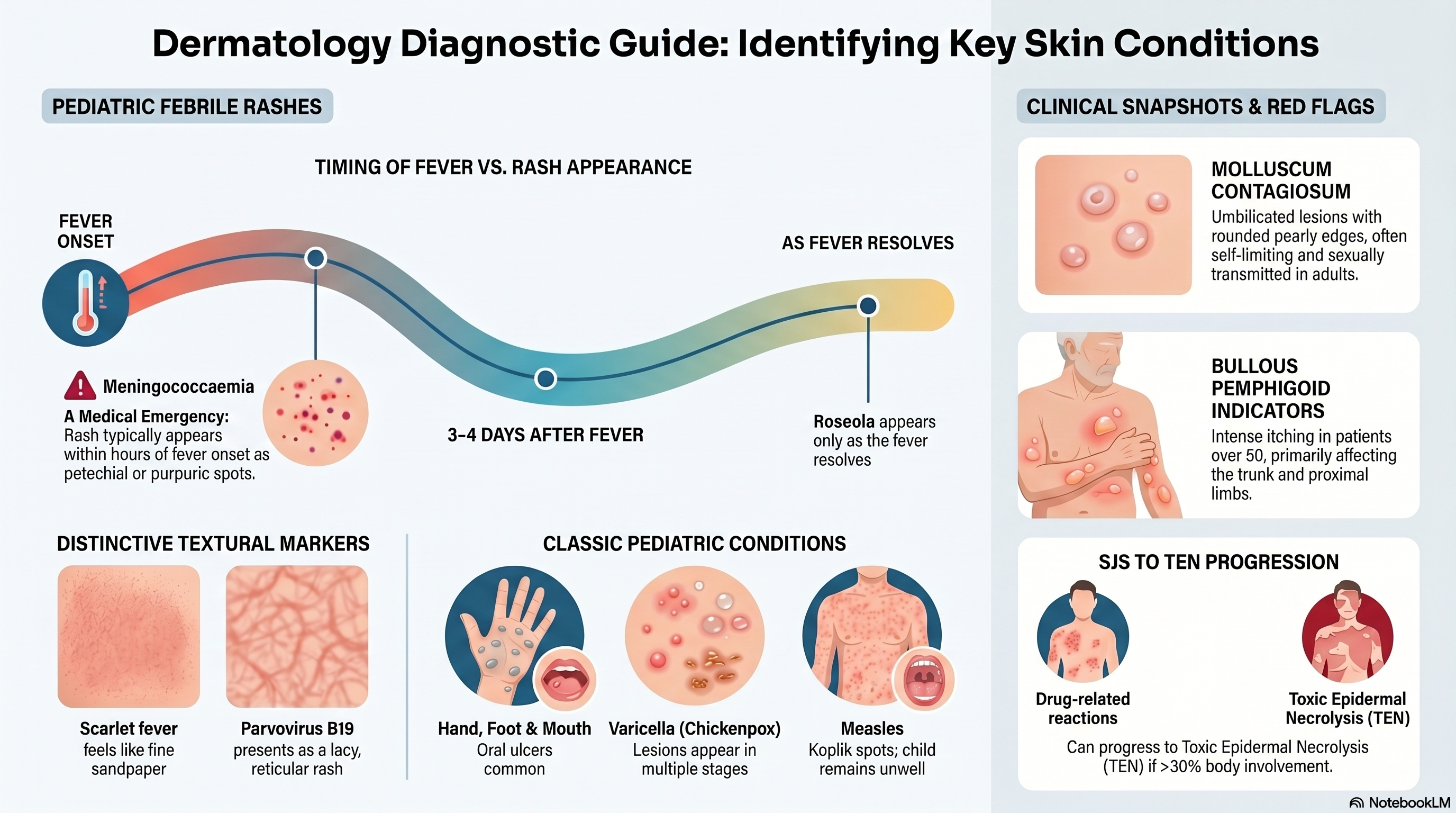
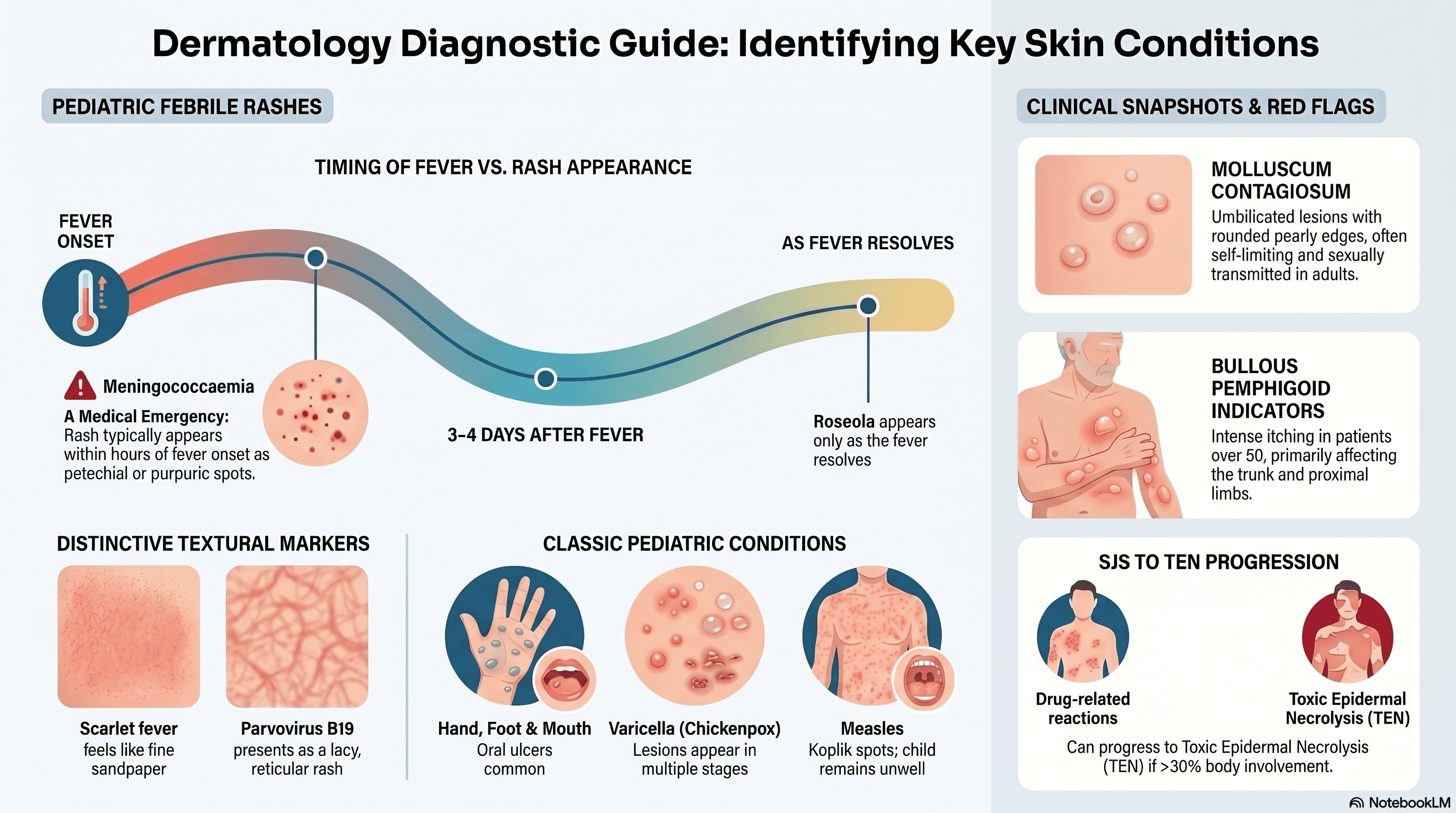 Dermatology Diagnostic Guide: Identifying Key Skin Conditions — PosterPDF · opens in a new tab
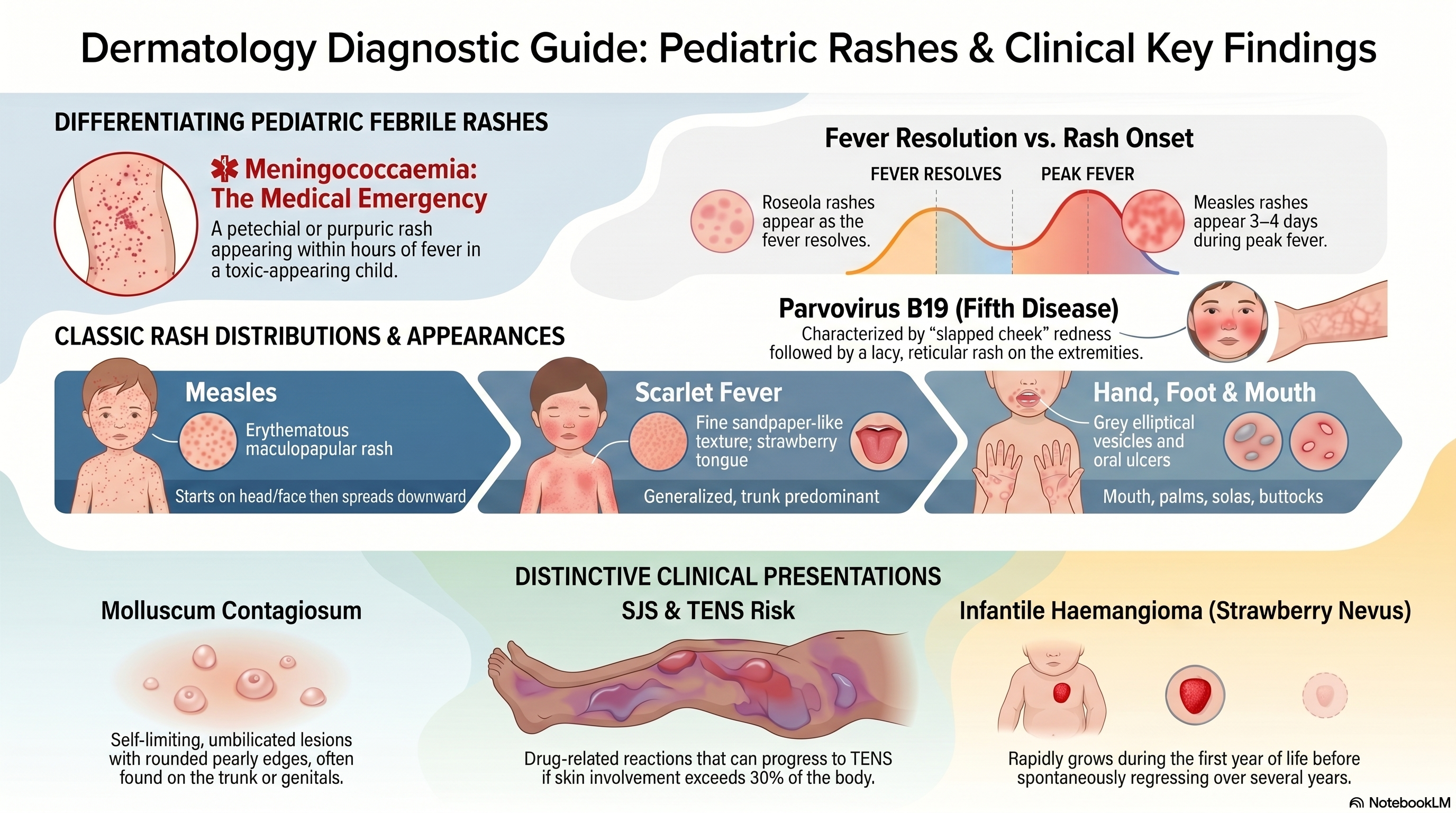
 Dermatology Diagnostic Guide: Paediatric Rashes & Clinical Key Findings — PosterPDF · opens in a new tab
Clinical pearls & learnings
Tips and tricks collected over the years — a living list that grows with every rotation. Use as a study guide only and check current guidelines before acting on anything.
Ulcers, cysts & pitted lesions
The painless foot ulcer — three to consider
| Cause | Key features |
|---|---|
| Venous ulcer | Gaiter area (medial malleolus), irregular/shallow, associated oedema, hemosiderin staining and varicosities |
| Buruli ulcer (Mycobacterium ulcerans) | Endemic to parts of coastal Victoria and northern Australia; starts as a painless nodule that breaks down into an ulcer with undermined edges — painless because the toxin (mycolactone) suppresses local immune response and damages nerves |
| Neuropathic ulcer (T2DM) | Over pressure points (plantar surface, metatarsal heads); painless due to loss of protective sensation from peripheral neuropathy |
Pitted keratolysis
A bacterial infection (Kytococcus sedentarius and related species) causing malodorous, punched-out pits on the sole, associated with hyperhidrosis and occlusive footwear. Treat with a topical antibiotic — mupirocin, clindamycin or fusidic acid — alongside moisture control (antiperspirants, breathable footwear, moisture-wicking socks).
Dermoid cyst
A dermoid cyst is a mature (benign) teratoma — a congenital cyst containing tissue from multiple germ layers (skin appendages, hair, sebaceous material, occasionally teeth or cartilage), typically found along embryonic fusion lines such as the lateral eyebrow, midline nose, or scalp.
Skin infections
Angular cheilitis
Often polymicrobial: Candida albicans (isolated in around 93% of cases) and Staphylococcus aureus (around 20%) frequently coexist, alongside Streptococcus species.
- Risk factors: type 2 diabetes, immunocompromise, older age, ill-fitting dentures, nutritional deficiency (iron, B12, folate, riboflavin), and a lip-licking/drooling habit that keeps the skin folds chronically moist.
Molluscum contagiosum
Dome-shaped, umbilicated papules with a pearly, rounded edge — classically across the trunk. Spread is by direct skin contact (including sexual contact in adults); in children, genital lesions are usually from autoinoculation/spread from other body sites rather than a marker of abuse, though the possibility should still be considered appropriately in context.
Self-limiting in immunocompetent patients — most resolve within 6–18 months without treatment, so deroofing/curettage isn't routinely needed. Active treatment is reserved for persistent, cosmetically bothersome, or immunocompromised cases.
Erythrasma
Corynebacterium minutissimum infection causing well-demarcated red-brown patches in skin folds (groin, axillae, toe webs) — fluoresces coral-pink under Wood's lamp (coproporphyrin III), a quick bedside way to distinguish it from tinea.
Treatment: topical fusidic acid first-line, oral or topical erythromycin second-line (or for more widespread disease).
Non-bullous impetigo
Choosing therapy
| Scenario | Treatment |
|---|---|
| 1–3 lesions, not in a high-risk group | Topical mupirocin ointment |
| Aboriginal and Torres Strait Islander or remote communities | Oral flucloxacillin preferred — higher burden of streptococcal disease and downstream complications (post-streptococcal glomerulonephritis, rheumatic fever) |
| Penicillin allergy | Oral cefalexin for a mild/non-severe allergy — use a non-beta-lactam alternative (e.g. trimethoprim-sulfamethoxazole, clindamycin) if the allergy history is severe/anaphylactic |
Dissecting cellulitis of the scalp
Also called perifolliculitis capitis abscedens et suffodiens — part of the follicular occlusion tetrad alongside hidradenitis suppurativa, acne conglobata and pilonidal disease. Painful nodules with interconnecting sinus tracts and purulent discharge.
- Oral doxycycline (± rifampicin) for milder disease.
- Intralesional corticosteroid injections for inflammatory nodules.
- Oral isotretinoin where there's scarring, keloid formation or patchy alopecia.
- Biologics (e.g. adalimumab) are an emerging option for refractory disease.
Inflammatory & genital dermatoses
Perioral dermatitis
Remove the trigger — classically topical corticosteroid use (even low-potency), and heavy/occlusive moisturisers or cosmetics — plus oral doxycycline for 6–12 weeks. Expect an initial flare after stopping steroids before it improves; topical metronidazole or erythromycin can be used for milder disease.
Lichen sclerosus (genital)
In males, the genital form is also called balanitis xerotica obliterans (BXO) — white, atrophic patches causing a tight foreskin that can't be fully retracted (phimosis), which can progress to urethral meatal stenosis and a deviated or spraying urinary stream. Circumcision is often required where phimosis is significant.
In females, lichen sclerosus classically causes itchy, atrophic white plaques in a "figure-of-8" distribution around the vulva and perianal skin, with a small increased long-term risk of vulval squamous cell carcinoma — worth long-term follow-up.
Guttate psoriasis
Small, "drop-like" papules over the trunk and proximal limbs, classically triggered by a preceding infection — usually streptococcal (e.g. tonsillitis). May resolve spontaneously over weeks to months, though a subset go on to develop chronic plaque psoriasis.
Vascular & bullous conditions
Infantile haemangioma (strawberry naevus)
Usually absent or minimal at birth, appearing in the first few weeks of life, then growing rapidly through the first year (proliferative phase) before spontaneously regressing over the following years (involution) — this growth-then-regression pattern is what distinguishes it from a vascular malformation, which is present at birth and grows proportionately with the child without regressing.
Most need no treatment. Oral propranolol is first-line where treatment is indicated — ulceration, visual axis or airway compromise, or a cosmetically significant facial lesion.
Stevens-Johnson syndrome / toxic epidermal necrolysis (SJS/TEN)
Classified by body surface area with epidermal detachment
| Entity | BSA detached |
|---|---|
| SJS | <10% |
| SJS/TEN overlap | 10–30% |
| TEN | >30% |
Classic culprits: allopurinol, anticonvulsants (carbamazepine, lamotrigine, phenytoin), sulfonamides, and NSAIDs.
Stop the culprit drug immediately and manage as a medical emergency — patients need burns unit/ICU-level supportive care given the risk of sepsis, fluid loss and multi-organ involvement.
Bullous pemphigoid
An autoimmune subepidermal blistering disease — typically older patients, intensely itchy, tense bullae on an erythematous/urticated base, mainly the trunk and proximal limbs. Mucous membranes are usually spared, and the bullae are tense (unlike pemphigus vulgaris, which more often involves mucosa and has flaccid, easily ruptured blisters) — a useful bedside differentiator.
Paediatric fever & rash
Erythema toxicum neonatorum
A benign, self-limiting neonatal rash requiring no treatment beyond parental reassurance — typically appears within the first few days of life and resolves spontaneously within 1–2 weeks.
Fever + rash — sorting the differential
| Condition | Rash timing vs fever | Distribution | Classic appearance | Key distinguishing features |
|---|---|---|---|---|
| Measles | 3–4 days after fever onset | Head/face → spreads downward | Erythematous maculopapular | Child remains unwell; Koplik spots |
| Roseola infantum (HHV-6/7) | After 3–5 days of fever, as fever resolves | Trunk first, then neck & proximal limbs | Fine pink macules/papules | Well child when rash appears |
| Rubella | Same time or shortly after fever | Face → trunk → extremities | Fine maculopapular | Lymphadenopathy (post-auricular/suboccipital), mild illness |
| Parvovirus B19 | After systemic symptoms resolve | Cheeks → arms & legs | "Slapped cheek", then lacy reticular rash | Nasal ridge & perioral sparing; no longer infectious once rash appears |
| Enterovirus | Late in illness | Variable | Non-specific maculopapular | Often mild viral features |
| Scarlet fever (GAS) | 12–48h after fever onset | Generalised, trunk predominant | Fine sandpaper-like rash | Strawberry tongue, circumoral pallor, Pastia's lines in skin folds |
| Hand, foot and mouth disease | With or shortly after fever onset | Mouth, palms, soles, buttocks | Grey elliptical vesicles | Oral ulcers common |
| Varicella (primary — chickenpox) | 24–48h after prodrome | Trunk & face first → periphery | Papules, vesicles, pustules & crusts together | Lesions in multiple stages at once ("crops") |
| Varicella (secondary — shingles) | ± fever | Dermatomal, unilateral | Clusters of vesicles on erythematous base | VZV reactivation |
| Kawasaki disease | During febrile phase (≥5 days fever) | Polymorphous, often trunk/perineum | Polymorphous, non-vesicular rash | Mucocutaneous changes — conjunctivitis, red/cracked lips, strawberry tongue, extremity changes, cervical lymphadenopathy |
| Meningococcaemia | Within hours of fever onset | Trunk & limbs | Petechial or purpuric, non-blanching | Toxic-appearing child — medical emergency |
| Gianotti-Crosti syndrome | Variable, post-viral | Extensor limbs, buttocks, face; trunk spared | Papular or papulovesicular | Symmetrical eruption, child often well |
Two diagnoses on this list must never be missed. Kawasaki disease untreated carries a real risk of coronary artery aneurysm — treat (IVIG + aspirin) on clinical grounds without waiting for a confirmatory test. Meningococcaemia with a non-blanching rash and fever is a medical emergency — same-day assessment and IV antibiotics without delay.