Neurology
Stroke, seizures, headache, neuro exam.
Use as a study guide only. These notes are part of a free open-access medical education (FOAMed) project and may contain errors or outdated information. Always verify against current guidelines (e.g. eTG, RACGP, local health district policies) and reputable sources before applying anything to patient care. See the full disclaimer.
 NeurologyPDF · opens in a new tab
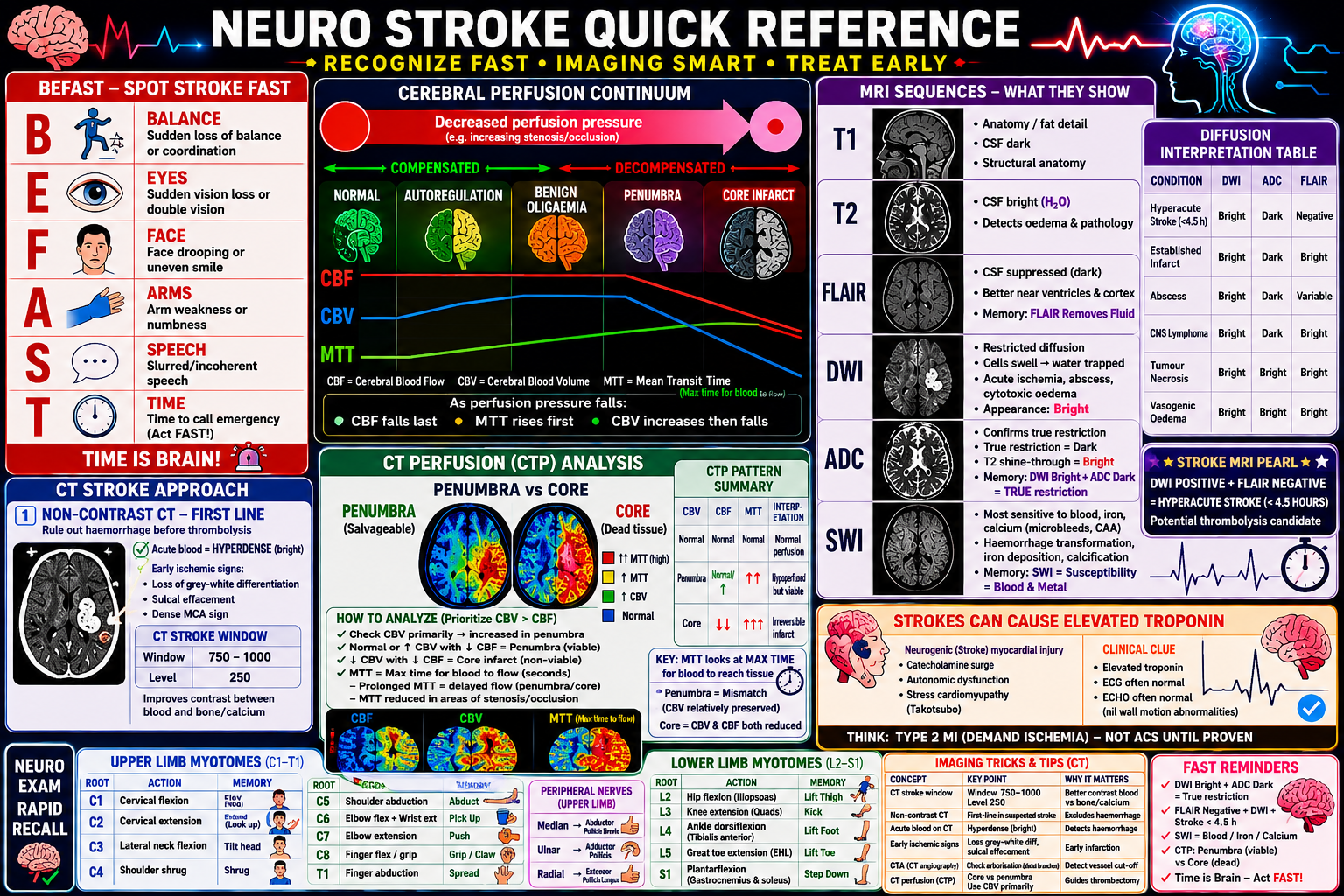
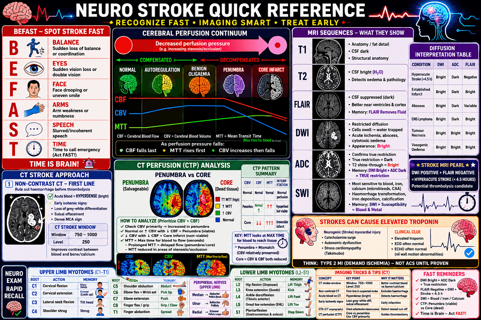 Neuro Stroke Quick Reference — PosterPDF · opens in a new tab
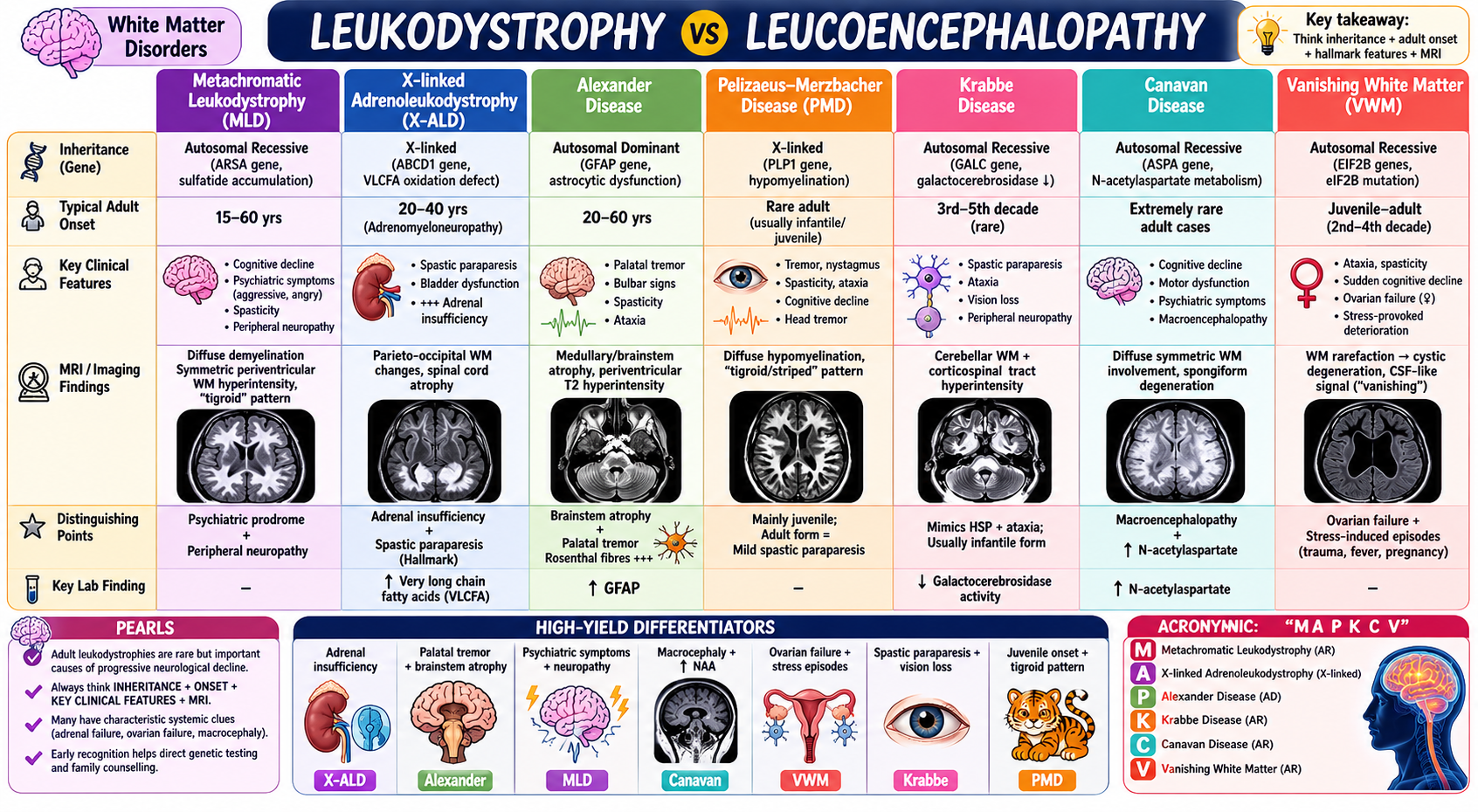
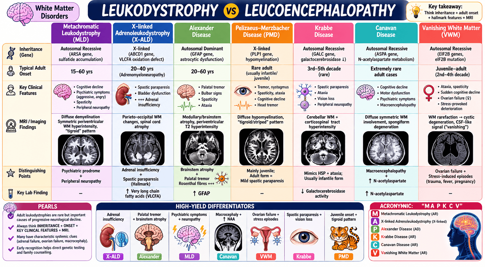 Leukodystrophy vs Leucoencephalopathy — PosterPDF · opens in a new tab
Clinical pearls & learnings
Tips and tricks collected over the years — a living list that grows with every rotation. Use as a study guide only and check current guidelines before acting on anything.
Headache
Thunderclap headache — think RCVS
Reversible Cerebral Vasoconstriction Syndrome classically causes recurrent thunderclap headaches over days to weeks. Imaging can be normal early — the absence of an infarct doesn't exclude it — but it can progress to ischaemic stroke (often watershed territory), convexity SAH, or PRES-like vasogenic oedema, sometimes presenting with a new focal neurological deficit.
- Triggers: vasoactive/serotonergic substances (SSRIs, triptans, nasal decongestants, cannabis, other sympathomimetics), the postpartum period, and stress.
- Differential for the same clinical picture: aneurysmal SAH (always exclude first), primary angiitis of the CNS, migraine, idiopathic intracranial hypertension, mass lesion, and MS.
- Imaging: CT angiogram may show multifocal segmental vasoconstriction ("string of beads"); a localised convexal SAH (confined to a few sulci, not the basal cisterns) is a recognised RCVS finding. Catheter (digital subtraction) angiography remains the gold standard when the diagnosis is unclear.
Management: a calcium channel blocker (oral/IV nimodipine, or verapamil) can reduce headache frequency and severity — watch for hypotension, which may need vasopressor support. Remove any triggering agent. Most cases resolve spontaneously within about 3 months.
The unusual headache — keep structural causes on the list
- A well-demarcated extra-axial lesion — think meningioma. Intra-axial possibilities include abscess, cyst, and stroke; also consider hydrocephalus.
- Any patient with a VP shunt and a new headache: consider both shunt malfunction and an abdominal CSF pseudocyst — a recognised complication (roughly 1–5% of shunts) where CSF collects around the peritoneal catheter tip within a fibrous capsule.
- Cerebral venous sinus thrombosis in a patient with inflammatory bowel disease — IBD is a prothrombotic state, especially with active disease, and CVST needs to be actively excluded in this group.
Trigeminal neuralgia
Brief, severe, electric-shock-like facial pain in a trigeminal distribution, often triggered by light touch. Atypical features — younger age, bilateral pain, or sensory loss — should prompt MRI to exclude a secondary cause: stroke, mass effect from a tumour, or a vascular malformation/compressive vessel.
First-line medical therapy is carbamazepine (or oxcarbazepine); gabapentin, pregabalin and lamotrigine are alternatives or adjuncts. Refractory or secondary cases may need surgical options such as microvascular decompression.
Migraine & cluster headache prophylaxis
Match the prophylactic to the headache
| Indication | Agent | Caveat |
|---|---|---|
| Migraine prophylaxis | Propranolol (or another beta-blocker) — a mainstay | Avoid in asthma/reactive airway disease — use amitriptyline, topiramate or candesartan instead |
| Cluster headache prophylaxis | Verapamil — the mainstay specifically for cluster | — |
| Chronic migraine (≥15 headache days/month) | Botulinum toxin — approved for this indication | — |
| Refractory migraine and cluster | Sphenopalatine ganglion block — 2% lignocaine, roughly 2.5 mL into each nostril | Bathes the region of the ganglion in local anaesthetic |
Stroke
Stroke can raise troponin — don't chase a false ACS
Neurogenic (type 2) myocardial injury from a catecholamine surge and autonomic dysfunction can raise troponin after an acute stroke, sometimes with a stress-cardiomyopathy (Takotsubo) picture. The ECG and echo are often unremarkable — no significant wall motion abnormality — but don't assume this every time; a genuine concurrent ACS is still possible and should be considered on its own merits.
Always document the precise stroke territory/location at handover — vague descriptions cost the next team time when they're assessing for a new deficit.
Stroke while anticoagulated
- Check an anti-factor Xa level if the patient is on a factor Xa inhibitor (apixaban, rivaroxaban) — this tells you whether they were actually therapeutically anticoagulated, which matters for both the likely mechanism and thrombolysis eligibility decisions.
- TTE (± TOE for higher sensitivity) to look for a cardioembolic source — LV or atrial thrombus, valvular disease, PFO.
- Review adherence and dosing, and consider whether a different agent or dose is warranted — don't assume the drug "failed" without checking these first.
Haemorrhagic stroke (SAH) — acute management
- Stop and reverse any anticoagulant/antiplatelet
- Head of bed elevated to 30°
- Control blood pressure (SBP roughly 140–160, or per local protocol) — labetalol first-line, hydralazine as an alternative
- Nimodipine to reduce vasospasm/delayed cerebral ischaemia (standard in aneurysmal SAH)
- CT angiogram to define the vascular anatomy, then neurosurgical/interventional discussion — aneurysm: coiling or clipping; AVM: embolisation, resection or radiosurgery depending on grade
Be cautious with mannitol in SAH specifically — maintaining euvolaemia (not aggressive diuresis) helps prevent delayed cerebral ischaemia from vasospasm. It still has a role for impending herniation, but isn't used routinely here.
Posterior circulation stroke vs cerebral venous sinus thrombosis
Posterior circulation stroke, central vertigo and CVST can all present similarly — vertigo, headache, cerebellar signs. Look specifically for CVST features: a severe, worsening headache, new seizures, or focal neurological signs.
Imaging for suspected CVST
| Test | Sign | Note |
|---|---|---|
| Non-contrast CT | "Dense triangle" sign — hyperdense thrombus in the superior sagittal sinus | Recognised but insensitive — a normal CT does NOT exclude CVST |
| CT venogram | "Empty delta" sign — a central filling defect in the sinus surrounded by enhancing collaterals/dura | — |
| MRI + MR venography | — | The preferred non-invasive test where available |
Neuro exam signs
Frontal release signs
Primitive reflexes that re-emerge with frontal lobe dysfunction — a frontal lobe lesion (stroke, trauma, tumour), or diffuse processes like dementia, Parkinson's disease, or encephalopathy.
- Grasp, rooting and sucking reflexes.
- Palmomental reflex — stroking the thenar eminence causes contraction of the ipsilateral chin/mentalis muscle.
- Glabellar tap (Myerson's sign) — repetitive tapping on the glabella normally causes blinking that quickly habituates; persistent blinking that fails to habituate is classically associated with Parkinson's disease.
New UMN signs with back pain = cord compression until excluded
New bilateral leg weakness, hyperreflexia or a sensory level in a patient with back pain is spinal cord compression until proven otherwise — epidural abscess, malignancy, disc prolapse. This needs an emergency MRI of the whole spine and urgent surgical/oncological input.
An upgoing (extensor) plantar response can be the earliest sign of an evolving UMN lesion, sometimes preceding other findings — don't dismiss it while other signs are still developing.
Finger tapping: Parkinsonian bradykinesia vs a new stroke
On finger tapping/finger fractionation testing, a progressive decrement in amplitude and speed with repetition is characteristic of Parkinsonian bradykinesia. A new stroke instead tends to cause a fixed reduction in speed and dexterity from the outset, without that progressive decrementing pattern.
Functional neurological disorder — supportive signs
- Symptoms and signs that don't localise to a single anatomical lesion, and are often inconsistent between examinations.
- Hoover's sign: weakness of hip extension on direct testing that resolves when the contralateral leg is actively flexed against resistance (involuntary hip extension) — a well-validated positive sign of functional leg weakness.
- A brisk crossed adductor reflex or palmomental reflex points toward organic UMN (pyramidal) pathology, which helps weigh against a purely functional presentation when the picture is ambiguous.
Blepharospasm — involuntary, often chronic contraction/blinking of the eyelids — is usually a primary focal dystonia (benign essential blepharospasm) rather than a functional disorder in its own right, though fatigue, stress and dry eyes can aggravate it.
Post-thrombolysis/thrombectomy monitoring: CT angiogram from the arch to the circle of Willis plus CT perfusion acutely, then a routine 24-hour non-contrast CT brain to check for haemorrhagic transformation.
Holmes–Adie pupil vs Argyll Robertson pupil
| Feature | Holmes–Adie pupil | Argyll Robertson pupil |
|---|---|---|
| Pupil size | Dilated, often unilateral | Small, irregular, usually bilateral |
| Speed of constriction | Slow, tonic | Rapid with near effort — accommodates but does not react to light |
| Reflexes | Reduced/absent deep tendon reflexes (classically ankle jerks) | Intact |
| Aetiology | Peripheral — postganglionic parasympathetic damage (ciliary ganglion) | Central — pretectal midbrain (classically neurosyphilis) |
| Typical patient | Young women, idiopathic or post-viral | Adults with tertiary syphilis (tabes dorsalis) |
| Prognosis | Usually benign | Pathological — treat the underlying syphilis |
Bulbar palsy vs pseudobulbar palsy
| Feature | Bulbar palsy | Pseudobulbar palsy |
|---|---|---|
| Site of lesion | LMN lesion of CN IX, X, XI, XII (medulla or peripheral nerve) | Bilateral UMN lesion of the corticobulbar tracts |
| Common causes | Motor neurone disease (progressive bulbar type), brainstem stroke, Guillain–Barré, myasthenia gravis, syringobulbia | Bilateral stroke, MS, motor neurone disease (UMN type), head trauma |
| Speech | Nasal, slurred — "flaccid dysarthria" | Slow, effortful — "spastic dysarthria" |
| Voice | Nasal, weak | Harsh, strained |
| Swallowing | Severe dysphagia, nasal regurgitation | Dysphagia, less nasal regurgitation |
| Tongue | Wasted, fasciculating, weak | Spastic, small, stiff — no wasting or fasciculations |
| Jaw jerk | Absent or weak | Exaggerated/brisk |
| Gag reflex | Absent | Exaggerated |
| Emotional control | Normal | Emotional lability (pathological laughing/crying) |
| Other CN findings | LMN signs — atrophy, fasciculations, palate weakness | UMN signs — brisk reflexes, spasticity, preserved bulk |
Demyelinating & inflammatory disease
NMOSD — a distinct diagnosis from MS
Neuromyelitis optica spectrum disorder is a separate antibody-mediated astrocytopathy, not a subtype of MS — the distinction matters because some MS disease-modifying therapies can worsen NMOSD. Classic pattern: optic neuritis (often severe, sometimes bilateral or simultaneous) plus longitudinally extensive transverse myelitis (≥3 vertebral segments on MRI), associated with anti-aquaporin-4 antibodies. A related but distinct entity, anti-MOG antibody disease (MOGAD), is now classified separately.
Transverse myelitis
Motor and sensory deficits below the lesion level plus autonomic (bladder/bowel) dysfunction. Get an urgent MRI spine to exclude a compressive cause (surgical emergency) before treating as inflammatory — causes include NMOSD, MOGAD, MS, para-/post-infectious myelitis, and idiopathic disease.
CADASIL
Cerebral Autosomal Dominant Arteriopathy with Subcortical Infarcts and Leukoencephalopathy — a NOTCH3 mutation causing migraine with aura, recurrent subcortical strokes, cognitive decline and mood disturbance. Management is mainly supportive: vascular risk factor control and genetic counselling, as there's no disease-modifying therapy.
Tumefactive MS
A large (>2 cm) demyelinating lesion that mimics a tumour on imaging, sometimes with mass effect and ring enhancement. Distinguishing it from neoplasm can be genuinely difficult radiologically, and biopsy is sometimes needed.
Reading the pattern: leptomeningeal vs white matter vs global encephalopathy
Pattern on imaging → what to think of
| Pattern | Finding | Causes |
|---|---|---|
| Leptomeningeal disease | Meningeal enhancement ± CSF pleocytosis | Infection, carcinomatous meningitis, sarcoidosis |
| White matter disease | T2/FLAIR hyperintensities | Demyelination, small vessel disease, leukodystrophy |
| Global encephalopathy | Diffuse cortical/subcortical dysfunction | Metabolic, toxic, infective, autoimmune or hypoxic causes |
Cortical ribboning (linear cortical hyperintensity following the gyral ribbon on DWI/FLAIR) is classically taught for Creutzfeldt-Jakob disease (prion disease), and also occurs with other causes of cortical laminar necrosis such as severe hypoxic-ischaemic injury or status epilepticus.
SUSAC syndrome
A rare autoimmune endotheliopathy affecting the microvasculature of the brain, retina and inner ear — the triad of encephalopathy, branch retinal artery occlusion (visual disturbance, scotomas) and sensorineural hearing loss. Differential includes MS and CNS vasculitis.
Peripheral neuropathy work-up
Working up a peripheral neuropathy
- Metabolic/nutritional screen: B12, folate, iron studies, thiamine (B1), B6, vitamin D, vitamin E. Add serum caeruloplasmin/24-hour urinary copper and zinc where there's a relevant risk factor for copper deficiency (e.g. prior bariatric surgery, malabsorption, high-dose zinc supplementation) rather than as a routine screen for everyone.
- Infective causes: HIV and syphilis (tabes dorsalis) directly cause neuropathy; also consider recent infection as a Guillain–Barré trigger (Campylobacter, EBV, CMV, Zika) where the pattern is acute and ascending.
- MRI brain and spine if a central cause or myelopathy/radiculopathy mimic needs excluding.
- Nerve conduction studies/EMG to characterise demyelinating vs axonal pathology and localise the lesion.
Movement disorders
Comparing the abnormal movements
| Movement | Description | Classic cause/exclude by |
|---|---|---|
| Tremor | Rhythmic, oscillatory movement around a joint axis | Resting (Parkinson's, 4–6 Hz, improves with action), postural (essential tremor, improves with alcohol), or intention (cerebellar disease, worsens approaching target) |
| Dystonia | Sustained or intermittent muscle contraction causing abnormal, often repetitive twisting movements or postures | Primary/genetic, drug-induced (acute dystonic reaction — antipsychotics), or a structural basal ganglia lesion |
| Athetosis | Slow, writhing, continuous movements — especially fingers and hands | Basal ganglia injury in infancy (kernicterus, hypoxic-ischaemic encephalopathy) — often coexists with chorea as "choreoathetosis" |
| Chorea | Rapid, irregular, non-rhythmic, flowing movements that seem to migrate between body parts | Huntington's disease, Sydenham's chorea (post-streptococcal), drug-induced (levodopa, tardive dyskinesia), lupus, thyrotoxicosis |
| Myoclonus | Sudden, brief, shock-like jerks — positive (contraction) or negative (sudden loss of tone, e.g. asterixis) | Metabolic encephalopathy (uraemia, hepatic failure), myoclonic epilepsy, neurodegenerative disease (CJD), post-hypoxic injury |
| Tics | Sudden, repetitive, stereotyped movements or vocalisations, preceded by an urge and briefly suppressible | Tourette syndrome (motor + vocal, childhood onset) |
Rhythmicity and suppressibility are the two fastest sorting questions: tremor is rhythmic (nothing else on this list is); tics are the only one that's voluntarily suppressible, at the cost of a building urge.
Huntington's disease
- Autosomal dominant (CAG repeat expansion, with anticipation) — always check family history and involve genetic counselling.
- Psychiatric and cognitive change often precedes or accompanies the onset of chorea — this is not purely a movement disorder.
- Classic exam finding: the "jack-in-the-box" tongue — inability to sustain tongue protrusion, darting in and out involuntarily (motor impersistence).
Neuroleptic malignant syndrome vs serotonin syndrome
| Neuroleptic malignant syndrome | Serotonin syndrome | |
|---|---|---|
| Trigger | Excess dopamine receptor antagonism (antipsychotics) or abrupt withdrawal of a dopaminergic drug (e.g. stopping levodopa) | Serotonergic agent — often a new drug or dose increase |
| Tone | Lead-pipe (uniform) rigidity | Hyperreflexia, clonus, neuromuscular hyperactivity (especially lower limbs) |
| Other features | Markedly elevated CK, fever, autonomic instability | Fever, agitation, autonomic features |
| Onset | Over days | Typically within 24 hours of the causative agent |
Vertigo & BPPV
Diagnosing BPPV
- A positive Dix-Hallpike shows positional nystagmus with a latency and fatigability, and no other neurological findings — non-fatigable nystagmus or additional neuro signs point to a central cause instead.
- Start the Dix-Hallpike with the side the patient is less symptomatic on to establish a baseline before testing the suspected side.
- Posterior canal BPPV (the most common form) produces upbeating, torsional nystagmus on Dix-Hallpike, with the affected ear being the one facing down.
Treating posterior canal BPPV — the Epley manoeuvre
- Start sitting, turn the head 45° toward the affected ear
- Lay the patient back quickly with the head extended slightly over the end of the bed (Dix-Hallpike position) — wait for nystagmus/dizziness to settle
- Turn the head 90° to the opposite side (now 45° away from the affected ear)
- Roll the body a further 90° so the patient is facing the floor
- Sit the patient up while keeping the head turned, then straighten
Horizontal canal BPPV
Diagnosed with the supine roll (Pagnini-McClure) test — geotropic nystagmus (beating toward the ground) is the more common canalithiasis variant, and the affected ear is the side that produces more intense nystagmus when turned.
- First-line treatment: the Lempert ("BBQ") roll — a sequence of 90° rolls totalling 360°, finishing sitting up.
- If that fails: the Gufoni manoeuvre — the patient lies quickly onto the unaffected side, the head is then turned 45° toward the floor and held, before sitting up.
Paediatric neurology
Raised ICP in infants
"Sunsetting" eyes — downward eye deviation with visible sclera above the iris, from pressure on the dorsal midbrain — is a classic sign of raised intracranial pressure in infants, alongside a bulging fontanelle, rapidly increasing head circumference, developmental delay or regression, vomiting and irritability. Think hydrocephalus, intracranial haemorrhage, or a mass lesion.