Haematology
Anaemia, clotting, leukaemia, transfusion.
Use as a study guide only. These notes are part of a free open-access medical education (FOAMed) project and may contain errors or outdated information. Always verify against current guidelines (e.g. eTG, RACGP, local health district policies) and reputable sources before applying anything to patient care. See the full disclaimer.
 Haematology & OncologyPDF · opens in a new tab
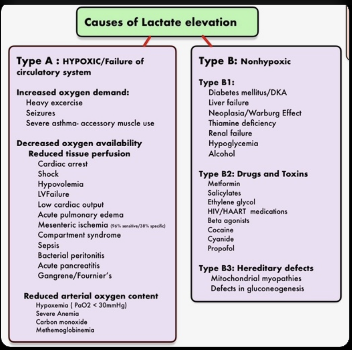
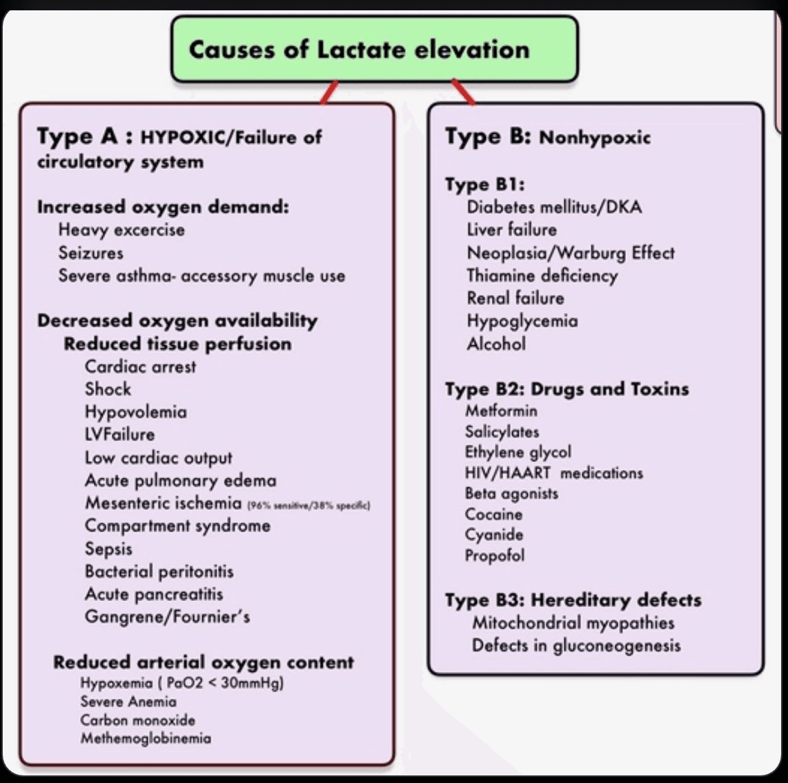 Causes of Lactate Elevation — PosterPDF · opens in a new tab
Clinical pearls & learnings
Tips and tricks collected over the years — a living list that grows with every rotation. Use as a study guide only and check current guidelines before acting on anything.
Anaemia & haemolysis
Mentzer index — microcytosis in one calculation
Mentzer index = MCV ÷ red cell count (RBC, ×10¹²/L). It helps separate the two commonest causes of microcytosis:
| Mentzer index | Suggests | Why |
|---|---|---|
| >13 | Iron deficiency anaemia | The marrow makes fewer cells, so RBC count falls proportionally more than MCV |
| <13 | Thalassaemia trait | The marrow makes plenty of small cells — RBC count is preserved or high despite a low MCV |
Note it is MCV divided by the red cell count, not by haemoglobin. It is a screening heuristic only — confirm with ferritin/iron studies and haemoglobin electrophoresis (and remember the two can coexist).
Always screen for haemolysis
- Core screen: LDH, haptoglobin, bilirubin (unconjugated), reticulocyte count, blood film, urate and CMP.
- Direct antiglobulin test (DAT/Coombs) to separate immune from non-immune causes.
- G6PD screen where the history fits — but check after the acute episode, since levels can be falsely normal during haemolysis when only young cells survive.
- Look for coexisting DIC: low fibrinogen with a high D-dimer.
Severe B12 deficiency can produce a strikingly high LDH — from ineffective erythropoiesis and intramedullary haemolysis inside the marrow. It can mimic a microangiopathic haemolytic anaemia, so check B12 before escalating to plasma exchange.
Pure red cell aplasia (PRCA)
- Isolated normocytic anaemia with profound reticulocytopenia and absent marrow erythroid precursors.
- Treatment: corticosteroids plus ciclosporin, then ciclosporin monotherapy for several months with therapeutic drug level monitoring.
- Screen for the underlying cause: thymoma (a well-recognised association — image the chest), parvovirus B19, lymphoproliferative disorders, and drugs.
Thymoma links PRCA to myasthenia gravis — treat myasthenia with pyridostigmine and immunotherapy, and consider thymectomy. (Hyperbaric oxygen has no role here.)
Raised lactate
Causes of a raised lactate
Not every lactate is sepsis.
Type A vs type B hyperlactataemia
| Type | Mechanism | Causes |
|---|---|---|
| Type A | Tissue hypoxia | Shock, ischaemia, severe hypoxaemia, profound anaemia |
| Type B | No tissue hypoxia | Liver failure, malignancy, thiamine deficiency, drugs and toxins (metformin, salbutamol, adrenaline, antiretrovirals), inborn errors of metabolism |
A lactate that will not clear despite adequate resuscitation should prompt two thoughts: is there ongoing ischaemia I have not found (mesenteric ischaemia, compartment syndrome, a missed source), and is this a type B cause — particularly thiamine deficiency, which is easy to treat and easy to miss.
Bleeding & coagulation
Interpreting the core bleeding panel
Pattern → cause → next test
| Pattern | Likely cause | Next investigation |
|---|---|---|
| Normal PT & aPTT with bleeding | Platelet function disorder or von Willebrand disease | vWF antigen + activity (ristocetin cofactor), factor VIII, platelet function assay (PFA-100) |
| Isolated ↑ aPTT | Haemophilia, heparin, lupus anticoagulant | Factor VIII / IX / XI assays; lupus anticoagulant, anti-β2GP1, anti-cardiolipin |
| Isolated ↑ PT | Warfarin, early vitamin K deficiency, liver disease, factor VII deficiency | Factor VII assay, vitamin K trial, LFTs |
| ↑ PT + ↑ aPTT + ↓ fibrinogen + ↓ platelets | DIC | High D-dimer with low fibrinogen — replace with cryoprecipitate/fibrinogen concentrate and treat the trigger |
| ↑ INR with normal platelets | Liver disease or warfarin | Factor V (low in liver disease, normal in vitamin K deficiency), LFTs, albumin |
The mixing study settles it: mix patient plasma 1:1 with normal plasma. Correction = factor deficiency (haemophilia, warfarin, liver disease, massive transfusion, vitamin K deficiency). Failure to correct = an inhibitor (lupus anticoagulant, acquired factor VIII inhibitor).
Heparin-induced thrombocytopenia (HIT)
- Immune-mediated and prothrombotic: antibodies form against platelet factor 4–heparin complexes, activating platelets.
- The problem is thrombosis, not bleeding — despite the low platelet count. Platelets typically fall >50% around days 5–10 of heparin exposure.
- Score the pre-test probability (4Ts), screen with anti-PF4 ELISA, and confirm with the serotonin release assay (highly specific).
- Stop all heparin (including flushes) and start a non-heparin anticoagulant — do not simply withhold anticoagulation, and avoid platelet transfusion.
Reversing anticoagulants
Agent → reversal
| Anticoagulant | Reversal | Caveat |
|---|---|---|
| Warfarin | Vitamin K plus prothrombin complex concentrate (Prothrombinex/Beriplex) for major bleeding | PCC works within minutes; vitamin K sustains the effect |
| Dabigatran | Idarucizumab — a true specific reversal agent | — |
| Factor Xa inhibitors (apixaban, rivaroxaban) | Andexanet alfa — a recombinant decoy factor Xa that binds the drug directly; PCC is the widely used alternative | Expensive, and carries a real thrombotic risk (VTE, ACS, stroke) because it also blunts endogenous anticoagulation |
Before reversing, check the anti-Xa level (drug-calibrated) and renal function — much of the decision hinges on whether the drug has already been renally cleared and when the last dose was taken.
Epistaxis — a stepwise approach
- Firm pressure over the soft anterior nares (not the bony bridge) for 10–15 minutes, sitting forward
- Topical vasoconstrictor — oxymetazoline (± topical local anaesthetic)
- Identify the bleeding point and cauterise — silver nitrate to one side of the septum only
- Anterior nasal packing; posterior packing and ENT referral if it continues
Consider tranexamic acid, and review anticoagulants and platelet count in anyone with recurrent or heavy bleeds.
Malignant haematology
Myeloma: watch for plasmacytoma
An extramedullary plasmacytoma — soft tissue swelling from a plasma cell mass — carries a worse prognosis in myeloma and responds less well to systemic therapy. Radiotherapy is the mainstay for local control, alongside systemic treatment.
Complications to keep on the radar
Complication → the test or move that catches it
| Complication | Setting | What to do |
|---|---|---|
| HLH (haemophagocytic lymphohistiocytosis) | New acute leukaemia with fevers and cytopenias | Check ferritin (strikingly elevated), triglycerides, fibrinogen, EBV status — treatable but rapidly fatal if missed |
| Cytokine release syndrome vs febrile neutropenia | Post-chemotherapy — both give fever and rising CRP | Timing and the agent given usually separate them — but treat for neutropenic sepsis first |
| ICANS (immune effector cell-associated neurotoxicity) | After CAR-T and bispecific antibodies | Screen with the ICE score for confusion, dysgraphia, reduced attention |
| CMV colitis | Immunosuppressed with diarrhoea | CMV PCR plus endoscopic biopsy |
| Docetaxel toxicity | Workhorse taxane in breast cancer | Watch for febrile neutropenia, fluid retention, peripheral neuropathy |
Antifungal and antiviral prophylaxis in AML is posaconazole plus an anti-herpes agent (valaciclovir/aciclovir). Bactrim (PJP prophylaxis) belongs to other regimens — for example ALL, and patients on prolonged high-dose steroids — not routine AML induction.
H. pylori — more than an ulcer
H. pylori is implicated in immune thrombocytopenia (ITP) and in gastric MALT lymphoma. Eradication alone can resolve both — test and treat before escalating to immunosuppression or chemotherapy.
Rare syndromes worth recognising
| Syndrome | Stands for | Recognise it by |
|---|---|---|
| VEXAS | Vacuoles, E1 enzyme, X-linked, Autoinflammatory, Somatic | Older men with unexplained inflammation, macrocytic anaemia, chondritis and vasculitis — somatic UBA1 mutation |
| POEMS | Polyneuropathy, Organomegaly, Endocrinopathy, Monoclonal protein, Skin changes | Paraprotein plus a progressive demyelinating neuropathy — often mislabelled as CIDP |
Rashes not to dismiss
- Leukaemia cutis — leukaemic infiltration of skin; implies systemic disease.
- Sweet syndrome (acute febrile neutrophilic dermatosis) — tender red plaques with fever; associated with malignancy (especially AML), infection and drugs.
- Pyoderma gangrenosum — rapidly enlarging ulcer with a violaceous undermined edge; do not debride.
- Graft-versus-host disease, panniculitis, and drug reactions — including beta-lactam rashes, which range from benign delayed maculopapular eruptions to severe cutaneous adverse reactions.
Most childhood “penicillin allergy” labels are viral exanthems or benign delayed rashes. Formal delabelling matters — carrying an incorrect label pushes patients onto broader, more toxic antibiotics.
Immunology & infection
Lip and facial swelling — allergy or bradykinin?
Histaminergic vs bradykinin-mediated angioedema
| Anaphylaxis / histaminergic | Bradykinin-mediated | |
|---|---|---|
| Skin | Urticaria, itch | No urticaria, no itch |
| Other features | Bronchospasm, hypotension | Slower onset |
| Responds to | Adrenaline, antihistamine, steroid | Does not respond to adrenaline or antihistamines |
| Think | Allergen exposure | ACE inhibitors; hereditary or acquired C1 esterase inhibitor deficiency |
| Treatment | Adrenaline first | C1-INH concentrate or icatibant |
Complement pattern
| C4 | C1-INH | C1q | |
|---|---|---|---|
| Hereditary angioedema (HAE) | Low | Low (type I) or dysfunctional (type II) | Normal |
| Acquired C1-INH deficiency | Low | Low | Low — the discriminator; look for lymphoproliferative disease |
Send mast cell tryptase at presentation and around 1, 4 and 8 hours, with a baseline level after recovery — a rise and fall supports anaphylaxis and helps when the diagnosis is contested later.
Returned traveller with fever
- Malaria first, always — thick and thin films (repeat ×3) or a rapid antigen test. It is the one that kills quickly.
- Then: dengue (NS1 antigen plus serology), chikungunya, typhoid (blood cultures), leptospirosis, rickettsial infection (look for an eschar), and hepatitis A and B.
- Take a proper exposure history: geography, dates, freshwater swimming, animal and insect bites, sexual contacts, food and prophylaxis taken.
Clostridioides difficile
Treatment by episode
| Episode | Treatment |
|---|---|
| First episode | Oral vancomycin 125mg QID for 10 days, or fidaxomicin. Metronidazole is now reserved for when neither is available, as it is less effective |
| First recurrence | Fidaxomicin, or a tapered and pulsed oral vancomycin course |
| Multiple recurrences | Tapered/pulsed vancomycin, fidaxomicin, faecal microbiota transplant, or bezlotoxumab |
Stop the precipitating antibiotic and any unnecessary PPI wherever possible.
Metronidazole is not recommended for recurrent C. difficile — recurrence is where vancomycin taper, fidaxomicin or FMT belong. Older teaching had this the other way around.
Transfusion & prescribing
Transfusion reactions
| Reaction | Clue | Immediate action |
|---|---|---|
| Febrile non-haemolytic | Isolated fever/rigors, otherwise well | Stop or slow; paracetamol; exclude the serious causes |
| Acute haemolytic (ABO incompatibility) | Fever, loin/back pain, dark urine, shock | Stop immediately, resuscitate, recheck identity, notify the blood bank |
| Allergic / anaphylaxis | Urticaria, wheeze, angioedema, hypotension | Stop; antihistamine for mild; adrenaline, hydrocortisone and oxygen for anaphylaxis |
| TACO (circulatory overload) | Hypertension, raised JVP, APO — commoner than you think | Stop, sit up, oxygen, furosemide |
| TRALI | Hypoxia and bilateral infiltrates within 6 hours, usually normotensive | Stop, respiratory support — diuretics do not help |
TACO vs TRALI is the classic bind: TACO is hypertensive and fluid-overloaded and improves with diuresis; TRALI is a permeability oedema that does not.
Prescribing conversions — use with care
- Steroids: dexamethasone is roughly 6–7× as potent as prednisolone — dexamethasone 0.75mg ≈ prednisone/prednisolone 5mg (so 1mg dexamethasone ≈ 6.5mg prednisolone). Dexamethasone has minimal mineralocorticoid effect and a much longer duration.
- Opioids: anchor conversions on oral morphine equivalents (IV fentanyl 100mcg ≈ IV morphine 10mg ≈ oral morphine 30mg ≈ oral oxycodone ~20mg), then reduce the calculated dose by 25–50% for incomplete cross-tolerance.
Opioid equianalgesic tables vary widely between sources and none account for the individual patient. Always use your local guideline or an approved calculator, reduce for cross-tolerance, and prescribe breakthrough analgesia rather than converting from memory.
Trimethoprim–sulfamethoxazole (Bactrim) in the elderly
- It is a CYP2C9 inhibitor, not an inducer — it raises the levels of the drugs it interacts with. The classic trap is a sharp rise in INR in a warfarinised patient.
- Also interacts with phenytoin, sulfonylureas (hypoglycaemia) and methotrexate.
- Trimethoprim blocks ENaC, causing hyperkalaemia, and competitively inhibits creatinine secretion — producing a rise in creatinine without a true fall in GFR.
In a frail older patient on warfarin, an ACE inhibitor or spironolactone, a short course of Bactrim can produce a supratherapeutic INR and dangerous hyperkalaemia together. Check INR and potassium a few days in, or choose a different agent.
Diabetes: pick the drug for the pattern
Match the agent to the glucose pattern
| Pattern | Agent | Trade-off |
|---|---|---|
| High post-prandial glucose | DPP-4 inhibitors (gliptins) — act on the incretin axis | Chiefly flattens post-meal peaks |
| High fasting glucose | Sulfonylureas (and basal insulin) | At the cost of hypoglycaemia and weight gain |
| Baseline therapy | Metformin remains first-line; add SGLT2 inhibitors or GLP-1 agonists early | Add early where there is cardiovascular, renal or weight indication |
HbA1c is uninterpretable within about 3 months of a blood transfusion — transfused donor cells distort the result. The same applies in haemolysis, recent major bleeding, haemoglobinopathies, iron deficiency and advanced CKD. Use fasting glucose or continuous monitoring instead.