Cardiology
Heart failure, ACS, arrhythmias, murmurs & ECGs.
Use as a study guide only. These notes are part of a free open-access medical education (FOAMed) project and may contain errors or outdated information. Always verify against current guidelines (e.g. eTG, RACGP, local health district policies) and reputable sources before applying anything to patient care. See the full disclaimer.
 CardiologyPDF · opens in a new tab
 ECG BasicsPDF · opens in a new tab
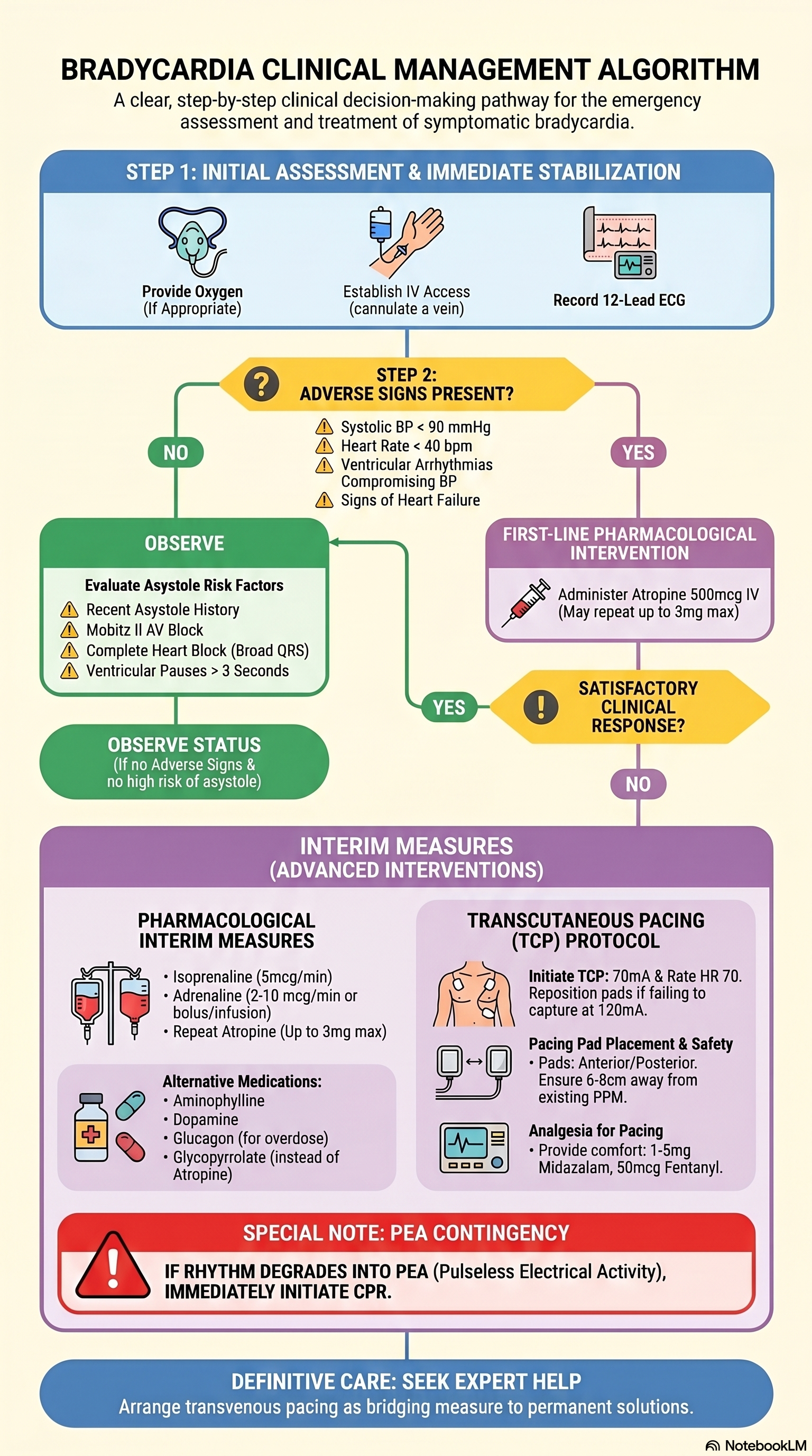
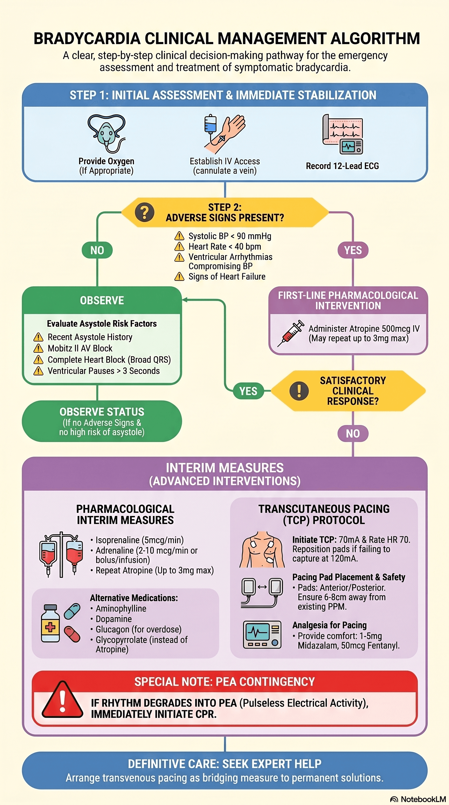 Bradycardia Clinical Management Algorithm — PosterPDF · opens in a new tab
- PACSA STEMI Reperfusion Flow Chart — PosterPDF · opens in a new tab
Clinical pearls & learnings
Tips and tricks collected over the years — a living list that grows with every rotation. Use as a study guide only and check current guidelines before acting on anything.
Risk stratification & ECG algorithms
Syncope and CVD risk tools
- San Francisco Syncope Rule — mnemonic CHESS: history of CHF, Haematocrit <30%, abnormal ECG, Shortness of breath, Systolic BP <90 at triage. Any one factor = high risk for a serious short-term outcome. Many EDs now use the Canadian Syncope Risk Score instead/alongside it — check your local protocol.
- For primary prevention decisions, use a validated absolute cardiovascular risk calculator (e.g. the Australian CVD risk calculator) rather than eyeballing individual risk factors.
Wellens syndrome — a warning of impending LAD occlusion
Deeply inverted or biphasic T waves in V2–V3 (± other precordial leads), seen during a pain-free interval, mark critical proximal LAD stenosis — Type A (biphasic) and Type B (deeply inverted, symmetric). These patients are at high risk of an extensive anterior MI if not intervened on, even with normal or minimally elevated troponin.
If chest pain recurs and the ECG reverts from the Wellens T-wave pattern back toward ST elevation, this suggests the LAD has occluded — repeat the ECG with any recurrence of pain rather than relying on a single strip.
The wide complex tachycardia
- By definition QRS >120ms; most VT sits in the 120–200ms range. An extremely wide QRS (>200ms) should make you think beyond typical VT — hyperkalaemia or sodium channel blocker toxicity (e.g. TCA overdose) can produce a very broad complex rhythm.
- Differential: VT, SVT with aberrancy (bundle branch block) or with pre-excitation, hyperkalaemia, severe ischaemia, a paced rhythm, sodium channel blockade, and fast AF with a bundle branch block.
- Treat as VT until proven otherwise, especially in a patient with known structural heart disease — assuming SVT with aberrancy in an unstable or ambiguous case is a common and dangerous error.
Sgarbossa criteria (and the modified/Smith criteria) help diagnose STEMI in the presence of LBBB or a paced rhythm, where standard ST-elevation criteria don't apply — look for ST elevation concordant with the QRS direction, or excessive discordant ST elevation (modified criteria: ST/S ratio ≥25%).
Conduction block terminology
- Bifascicular block: RBBB plus either left anterior fascicular block (LAFB) or left posterior fascicular block (LPFB) — this combination can progress to complete heart block.
- Trifascicular block: bifascicular block plus a prolonged PR interval (first-degree AV block affecting the remaining fascicle).
Prolonged QT
A prolonged QTc (not QRS) — roughly >450ms in men, >460ms in women, with >500ms carrying a substantially higher torsades risk. Causes include congenital long QT syndromes and drugs — classically TCA overdose, which also widens the QRS via sodium channel blockade. Management of TCA-related cardiotoxicity: IV sodium bicarbonate (overcomes the sodium channel blockade and narrows the QRS) plus IV magnesium for associated ventricular arrhythmia/torsades risk.
Acute coronary syndrome
STEMI and NSTEMI management
Reperfusion and antiplatelet strategy
| STEMI | NSTEMI | |
|---|---|---|
| Reperfusion | Primary PCI within 2 hours is the goal; where timely PCI isn't available, a pharmaco-invasive strategy with weight-based IV bolus tenecteplase | Coronary angiography — timing per risk stratification |
| Antiplatelets | Aspirin + ticagrelor (or another P2Y12 inhibitor) | Aspirin + clopidogrel or ticagrelor |
| Duration | Load DAPT in any ACS | DAPT typically ~12 months post-ACS, individualised against bleeding risk |
Post-MI complications — a rough timeline
| Timing | Complication |
|---|---|
| Earliest — first 24–48h especially | Ventricular arrhythmias (VT, VF) from electrical instability |
| First 1–2 weeks (commonly days 2–7) | Papillary muscle rupture → acute severe mitral regurgitation → sudden pulmonary oedema |
| First 1–2 weeks (commonly days 1–5) | Ventricular free wall rupture (tamponade) or septal rupture/VSD (biventricular failure) |
| Weeks later (classically 2–10 weeks) | Dressler syndrome — autoimmune pericarditis |
Keep spontaneous coronary artery dissection (SCAD) on the differential for AMI, especially in a younger patient — particularly a peripartum woman — without typical atherosclerotic risk factors. It's easily missed because the demographic doesn't fit the "usual" ACS profile.
Reperfusion injury after PCI/thrombolysis
Restoring flow to previously ischaemic myocardium can itself cause additional injury — reperfusion arrhythmias (including an accelerated idioventricular rhythm, generally benign and often taken as a marker of successful reperfusion), microvascular obstruction/no-reflow, myocardial stunning, and reperfusion oedema. Keep this in mind when the ECG or rhythm changes shortly after a STEMI is opened up — it doesn't necessarily mean re-occlusion.
Arrhythmia
New atrial fibrillation
- Look for a precipitant: infection, respiratory disease (pneumonia, PE), toxins, thyrotoxicosis, electrolyte derangement, rheumatic heart disease.
- Rate control example: metoprolol 12.5mg PO BD as a starting dose, titrated to response.
- Broadly: rate control (e.g. digoxin, beta-blockers) vs rhythm control (e.g. amiodarone) — the choice depends on duration, haemodynamic stability and patient factors. Digoxin has no role in a wide complex/ventricular arrhythmia — it's an AF-specific option.
Causes of bradycardia
- Sinus bradycardia
- Junctional rhythm
- Heart block (first, second — Mobitz I/II, or third-degree)
- Pacemaker malfunction
- Sick sinus syndrome
- Ectopic atrial rhythm
- Sinus exit block
TAVI and AV block
Transcatheter aortic valve implantation commonly disturbs the conduction system, which sits close to the annulus where the prosthetic valve is deployed — new AV block (sometimes complete heart block requiring a permanent pacemaker) is a recognised complication. Pre-existing conduction disease (baseline bradycardia, existing RBBB) is a strong predictor of this risk.
Aortic & vascular emergencies
Aortic dissection — clinical clues
- Radio-radial delay, or a systolic BP difference >20mmHg between arms (or between an arm and a leg), points to a dissection flap affecting a branch vessel — which arm/leg is affected depends on which branches the flap involves, not simply whether the dissection is ascending or descending.
- CT aortogram is the key investigation to define the extent and guide management.
Carotid artery stenosis
"Symptomatic" means a TIA or stroke referable to that carotid territory within the preceding 6 months — amaurosis fugax, contralateral limb weakness/sensory loss, or dysphasia (dominant hemisphere). Non-specific symptoms like headache, syncope or fatigue are not carotid territory symptoms and don't count towards this.
Intervention thresholds
| Stenosis | Approach | Note |
|---|---|---|
| Symptomatic ≥50% (particularly 70–99%) | Carotid endarterectomy preferred, ideally within 2 weeks of the qualifying event | Stenting is an alternative — favoured in high surgical/anaesthetic risk or anatomy unfavourable for surgery |
| Asymptomatic ≥70–80% | May be considered in selected patients with long life expectancy and low periprocedural risk | Benefit is smaller than for symptomatic disease — individualised; modern optimal medical therapy has narrowed the incremental benefit shown in older trials |
Nitrates — know the contraindications
Used for acute pulmonary oedema and ACS. Avoid in hypotension, and avoid in inferior MI with right ventricular involvement — the RV-infarcted heart is preload-dependent, and nitrates reduce preload, risking severe hypotension or cardiogenic shock.
Aortic stenosis — when leg oedema changes the picture
The classic severe-AS symptom triad is syncope, angina and dyspnoea — any of these warrants urgent workup for intervention. Peripheral (leg) oedema signals right heart failure/pulmonary hypertension from advanced disease, and carries the poorest prognosis — an urgent indication for valve intervention.
Hypertension — check both arms
A significant BP difference between arms, or a radio-radial delay, should prompt consideration of aortic coarctation or dissection rather than being dismissed as measurement error.
Fluids, drugs & perioperative cardiology
ACE inhibitor-induced angioedema
ACEi angioedema is bradykinin-mediated, not the IgE/histamine-mediated mechanism of typical allergic angioedema — it doesn't reliably respond to adrenaline or antihistamines. It can occur any time after starting the drug (including years later) and is an airway emergency if it involves the tongue, floor of mouth or larynx: secure the airway early, stop the ACEi permanently (and avoid ARBs, which carry a smaller but real cross-risk), and involve ENT/anaesthetics. Icatibant or FFP are specialist-guided options in severe cases.
Before you run in fluids
- Known heart failure — check a recent ECHO before committing to an aggressive fluid strategy.
- Acute pulmonary oedema — fluids will worsen it.
- AKI/CKD with oliguria or a rising creatinine — increased risk of fluid overload.
- Active bleeding or a coagulopathy — favour permissive hypotension over aggressive crystalloid, which dilutes clotting factors and can worsen bleeding.
Postural (orthostatic) hypotension — sorting the cause
Three broad mechanisms
| Mechanism | Causes |
|---|---|
| Volume / medication-related | Dehydration, prolonged bed rest/deconditioning, and drugs — alpha-blockers, nitrates, TCAs, dopamine agonists (e.g. levodopa), diuretics |
| Postprandial hypotension | After meals, from splanchnic blood pooling — seen with autonomic dysfunction (diabetes, Parkinson's disease, MND) |
| Chronic autonomic failure | Diabetes, Parkinson's disease, MND, CKD/dialysis, and aortic stenosis (fixed cardiac output limits the compensatory response) |
Work-up: review the medication chart, check a glucose, and get an ECHO if aortic stenosis is a possibility.
Delaying surgery after a cardiac event
Elective non-cardiac surgery is generally deferred for at least 3 months after an MI or coronary stent (longer where possible, and specifically until any mandatory dual antiplatelet therapy course is complete) — the perioperative risk of major adverse cardiac events (reinfarction, heart failure, death) is substantially higher in this early window.
Cardiac arrest & tamponade
Cardiac tamponade
Consider tamponade after chest trauma (blunt or penetrating) and after cardiac surgery or procedures — including pacemaker lead perforation and CABG.
Refractory VT/VF
- Antiarrhythmic options for VT/VF storm: amiodarone, lidocaine, or an esmolol infusion (beta-blockade is increasingly recognised for storm driven by sympathetic overdrive).
- Vasopressor support as needed — noradrenaline, adrenaline or metaraminol infusion depending on the haemodynamic picture.
- For refractory VF or pulseless VT, double sequential external defibrillation is an emerging technique with trial evidence of improved survival: two sets of pads (one antero-lateral, one antero-posterior) with shocks delivered in rapid succession per local protocol. Practical caveats: it requires two working defibrillators, and devices may need servicing/battery replacement afterward.
For refractory VF/VT, consider early transfer to a cath lab or ECPR-capable centre rather than prolonged on-scene resuscitation.